Assay & Application Notes
Automated Luminescence Cytotoxicity Assay: A Comparative Study
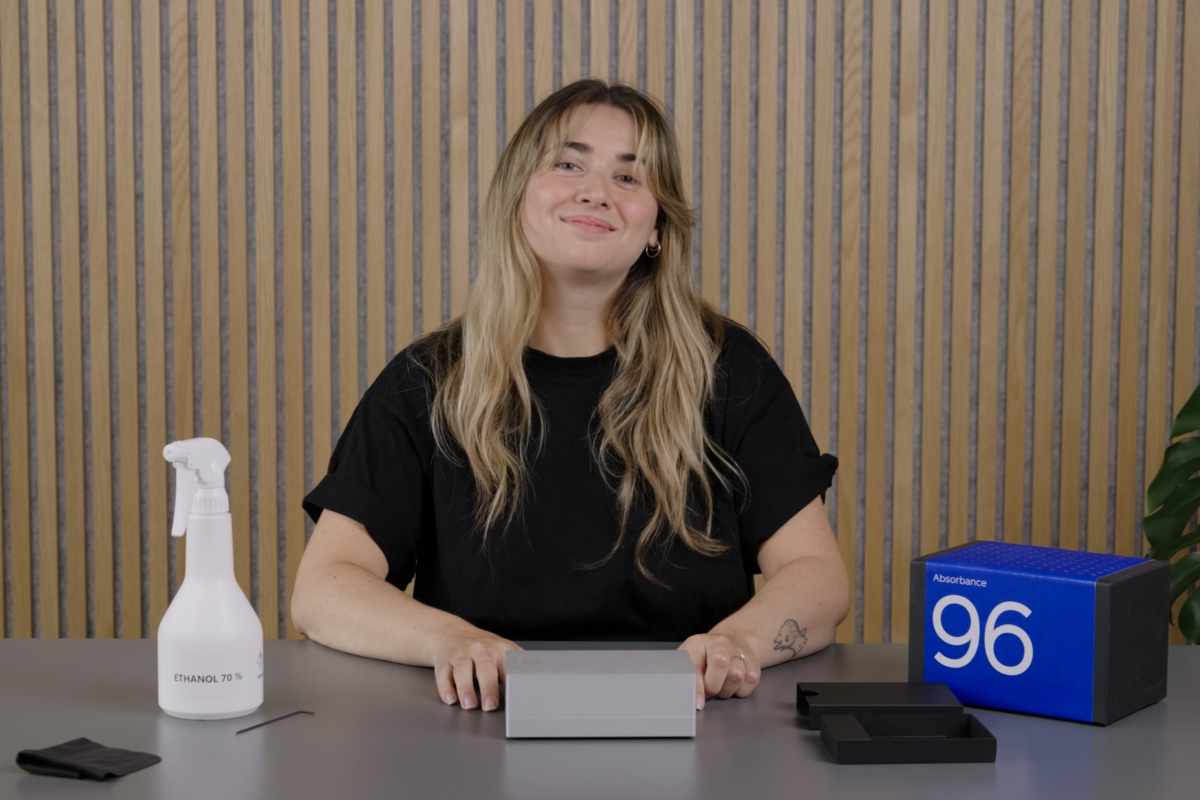
Integrating Luminescence 96 Automate with epMotion® liquid handling

Cytotoxicity assays are essential in various fields of biological research, drug discovery, and toxicology. They help assess cell viability and death in response to chemical, environmental, or biological stimuli. These assays are particularly useful for determining the toxic effects of new drug candidates, environmental agents, or other treatments on cellular systems. Luminescence-based assays, such as the CytoTox-Glo® assay, offer high sensitivity and ease of use, making them widely adopted for cytotoxicity measurements.
Automating Luminescence Assays with Integrated Luminescence Readout on Liquid Handling System
Automating the cytotoxicity assays streamlines the workflow (Fig. 1) by enabling precise, automated dispensing of reagents and samples and real-time luminescence detection. This integration significantly enhances data reproducibility by minimizing manual handling errors and reducing variability. The high-throughput capability, allowing up to 96 samples to be processed simultaneously, is particularly advantageous for large-scale cytotoxicity screenings. This workflow solution is an invaluable tool for drug discovery and cell biology researchers, where efficiency and reliability are critical.

Fig. 1: Workflow illustrating the steps involved in the cell-based assay using the CytoTox Glo™ Cytotoxicity Assay from Promega. The highlighted steps (blue) in the workflow can be performed using the epMotion® liquid handling system and the Luminescence 96 Automate.
When selecting a luminescence reader, key factors include sensitivity, which ensures the detection of low signal levels, and crosstalk; reduced crosstalk allows accurate measurement across different samples in the microplate. This study evaluated the performance and reliability of the Luminescence 96 Automate for automated luminescence assays and its integration with the epMotion® liquid handling system. The performance was benchmarked against a stand-alone plate reader from a competitor, comparing effectiveness in quantifying cell viability and cytotoxicity with CytoTox-Glo® assay by Promega. The goal was to establish an automated workflow that enhances precision and efficiency in high-throughput luminescence assays.
Results
Seamless Integration: Automating CytoTox-Glo® Cytotoxicity Assay with Luminescence 96 Automate and epMotion®
An automated CytoTox-Glo® cytotoxicity assay was conducted in triplicate to validate the integration of Luminescence 96 Automate into the epMotion® liquid handling system. The CytoTox-Glo® assay from Promega measures cell death by detecting protease activity released from cells with compromised membranes.
Adding 10 µl of the kinase inhibitor Staurosporine to the microplate induced its cytotoxic effect on the cells. Following the CytoTox-Glo® kit protocol, the luminescent signal (glow type) from dead cells was initially measured using Luminescence 96 Automate with a 0.3-second integration time. Subsequently, after adding the lysis reagent, the luminescent signal associated with the total number of cells was measured at the same integration time. By subtracting the luminescent signal resulting from experimental cell death from the total luminescent values, the viability (luminescent signal from live cells) was calculated. Additionally, for a comprehensive comparative analysis, signals were also measured using the stand-alone microplate reader from a competitor.

Fig. 2: Validation of Luminescence 96 Automate integration into the epMotion® liquid handling system using the CytoTox-Glo® cytotoxicity assay. Luminescent signals from dead cells were measured with Luminescence 96 Automate at a 0.3-second integration time (A), followed by a second measurement of total cell luminescence after lysis reagent addition (not shown). The respective Cell viability (B) was calculated by subtracting the luminescence of dead cells from total luminescence. For comparative analysis, measurements were also performed using the stand-alone microplate reader from a competitor (C, D). Results indicated comparable luminescent signal profiles between Luminescence 96 Automate and a stand-alone microplate reader from a competitor.
The results (Fig. 2) reveal comparable luminescent signal profiles between the Luminescence 96 Automate and the stand-alone microplate reader from a competitor.
Additionally, the half-maximal effective concentration (EC50) of Staurosporine, determined using a regression model fitted to a sigmoidal curve, showed consistent values using integrated and standalone luminescence reader (Fig.3) for both live and dead cell conditions (Fig. 3).

Fig. 3: Comparing the E50 value of Staurosporine for both live and dead cells also demonstrated similar values for Luminescence 96 Automate and stand-alone microplate reader from a competitor. EC50 values are in µM.
These findings demonstrate that the Luminescence 96 Automate effectively quantifies cell viability and cytotoxicity, producing data comparable to those obtained with another luminescence microplate reader. Importantly, its integration into the epMotion® system enables automated and immediate readouts without user intervention.
Conclusion
Comparative analysis ofLuminescence 96 Automate with a stand-alone microplate reader from a competitor confirmed consistent cell cytotoxicity quantification, highlighted by similar EC50 values across experimental conditions. Notably, its seamless integration into the epMotion® liquid handling system enables automated, immediate readouts without user intervention, setting it apart from traditional stand-alone plate readers with integration complexities for lab automation.
In conclusion, the established CytoTox-Glo® workflow significantly advances automated luminescence plate reading, offering researchers a powerful tool for conducting precise, high-throughput cellular luminescence assays confidently and efficiently.